
Presentation
Left-sided weakness, diplopia and right-sided facial palsy.
Patient Data


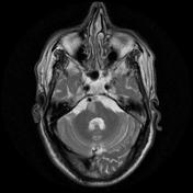
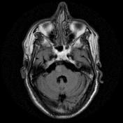

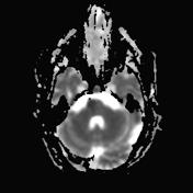
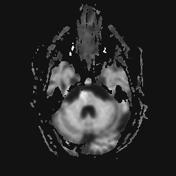
The right medial inferior pons shows a wedge-shaped area of restricted diffusion with high DWI and low ADC signal. It shows low T1 and high T2 / FLAIR signal intensity as well as subtle T1 hypointensity.
Multiple bilateral cerebral demyelinating foci with high T2/FLAIR signal intensity are seen with no restricted diffusion.
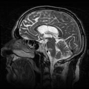
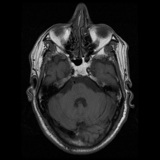
Mild dilatation of the ventricular system with peri-ventricular T2/FLAIR hyperintensity.
Widened extra-axial CSF spaces.
Normal sellar region. Normal cerebellum and cervico-medullary junction.
Case Discussion
This case shows typical right inferior medial pontine acute infarction; consistent with inferior medial pontine (Foville) syndrome. Atrophic brain changes with peri-ventricular arteriosclerotic leukoencephalopathy and bilateral cerebral small chronic vessel disease.
Inferior medial pontine (Foville) syndrome is characterized by:
- contralateral hemiparesis or hemiplegia due to corticospinal tract affection within the dorsal tegmentum of the pons at its inferior third.
- peripheral ipsilateral facial nerve palsy.
- conjugate gaze palsy with inability to look to the side of the lesion with diplopia.


 Unable to process the form. Check for errors and try again.