
Presentation
The onset of symptoms was left sided gaze palsy and left sided facial palsy. Underwent thrombolysis and symptoms regressed. MRI images were obtained 4 days after treatment.
Patient Data
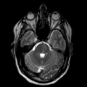

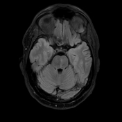
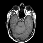
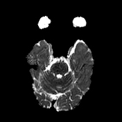

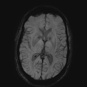
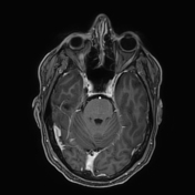

Subtle T2 hyperintense wedge-shaped area with signs of restricted diffusion located in the left inferomedial part of the pons. No clear sign of contrast enhancement.
Case Discussion
Isolated pontine infarcts account for 3% of all ischemic strokes and are caused mainly by atheromatous occlusive disease where the plaques protrude into the paramedian pontine branches, resulting in occlusion and infarction.
In this case, although the patient underwent thrombolysis 4 days prior to MRI image acquisition, areas of restricted diffusion were still present, contrary to the symptoms which regressed.


 Unable to process the form. Check for errors and try again.