
Presentation
Left groin pain and intermittent fullness with dysuria and hematuria.
Patient Data


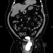
The left kidney is relatively small and shows parenchymal atrophic changes due to moderate to severe hydroureteronephrosis.
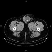
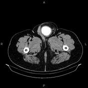
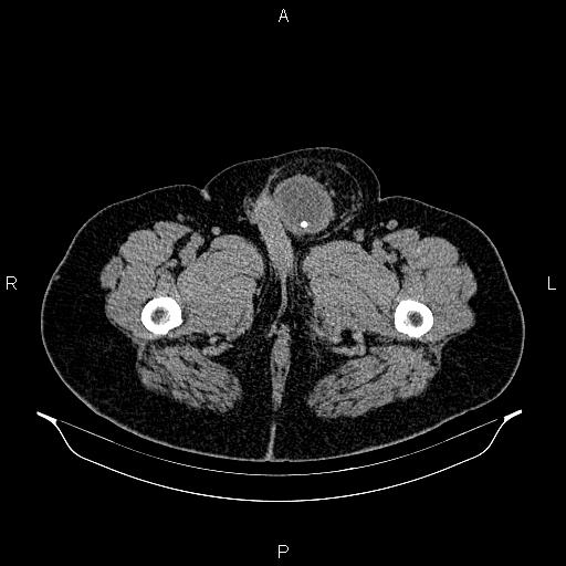
A sizable inguinal hernia is present on the left side, containing a large part of the urinary bladder and the distal part of the left ureter and causing obstruction. The herniated part of the urinary bladder shows wall thickening and surrounding fat stranding. Several stones are within the herniated bladder, less than 10 mm.
Case Discussion
This case demonstrates a left-sided inguinal hernia containing a large part of the urinary bladder and distal part of the left ureter that causes ipsilateral moderate to severe hydroureteronephrosis and left renal parenchymal atrophic changes.
It is important to be aware of this condition in any males older than 50 years with an inguinal hernia, as unknowingly, bladder injury during herniorrhaphy can lead to infection, sepsis, or death.


 Unable to process the form. Check for errors and try again.