
Presentation
Non-tender swelling along the left medial canthus since birth.
Patient Data
Age: 3 years
From the case:
Internal angular orbital dermoid cyst - fluid density


Download
Info
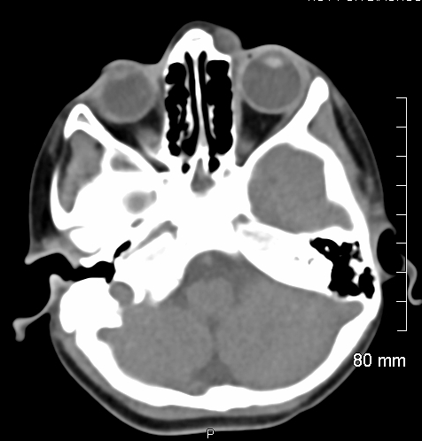
A small well-defined, thin walled, rounded cystic lesion measuring 9 x 8 mm, is seen in the preseptal compartment, along the medial canthus of the left orbit. It has an average density of 14 HU and shows no enhancing components, calcifications or intra-orbital extension. Smooth indentation and remodeling changes are seen in the underlying ethmoid bone.
From the case:
Internal angular orbital dermoid cyst - fluid density
Download
Info

Histopathology of the excised left orbital lesion lesion shows dermoid cyst.
Case Discussion
Small well-defined, cystic lesion along the left medial canthus which is likely an orbital dermoid cyst.


 Unable to process the form. Check for errors and try again.