
Presentation
Total hysterectomy with bilateral salpingo-oophorectomy for epidermoid carcinoma of the uterine cervix 2 months ago, presenting with swelling of the left abdomen without fever or significant abdominal pain. The ultrasound was inconclusive according to the radiologist. An MRI exam was requested by her surgeon.
Patient Data


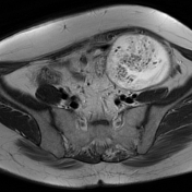
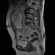
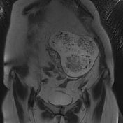
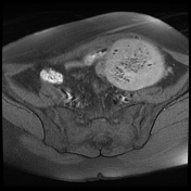
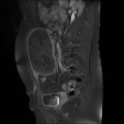
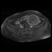
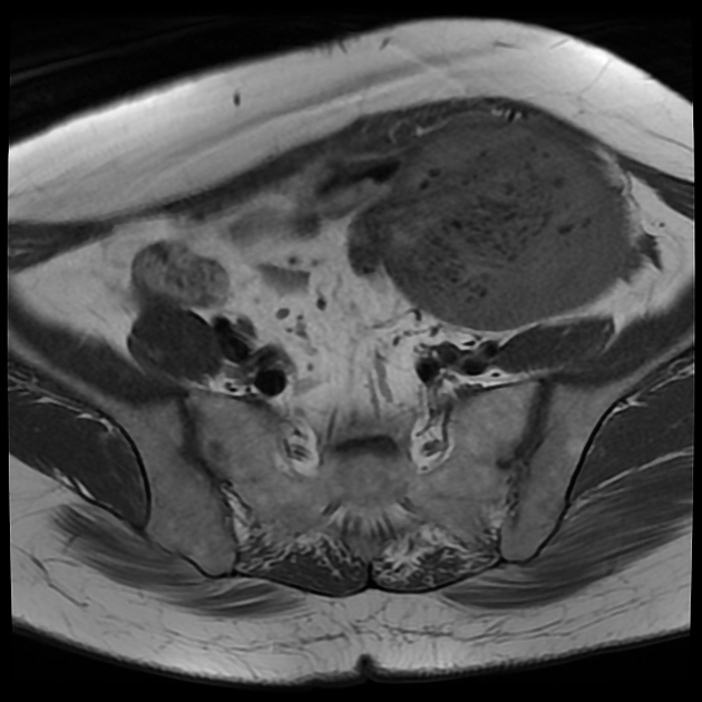
There is a large well-circumscribed left abdominopelvic mass of intraperitoneal location, of low signal intensity on T1WI, high signal intensity on T2WI, containing multiple gas-bubbles of low signal on all sequences. On postcontrast sequences, there is a peripheral rim enhancement. Note the coronal T2WI sequence shows like three contiguous masses with surrounding fluid collection.
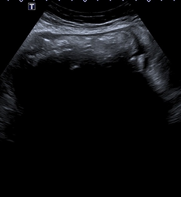
The ultrasound was performed after the MRI exam showing a complex echogenic mass with posterior acoustic shadowing, and surrounding fluid collection.
Case Discussion
The patient underwent an operation the next day and three gossypibomas (textilomas) were removed from the abdomen.
The ultrasound performed before the MRI exam did not mention the presence of an intrabdominal foreign body and the use of sponges with no radiopaque markers (as in this case) will be occult on plain radiograph.


 Unable to process the form. Check for errors and try again.