
Presentation
New onset of temporal lobe seizures and epilepsy.
Patient Data
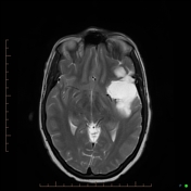

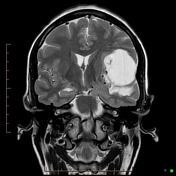

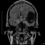
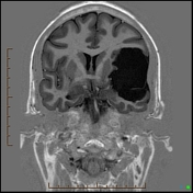
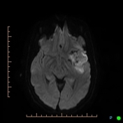
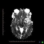
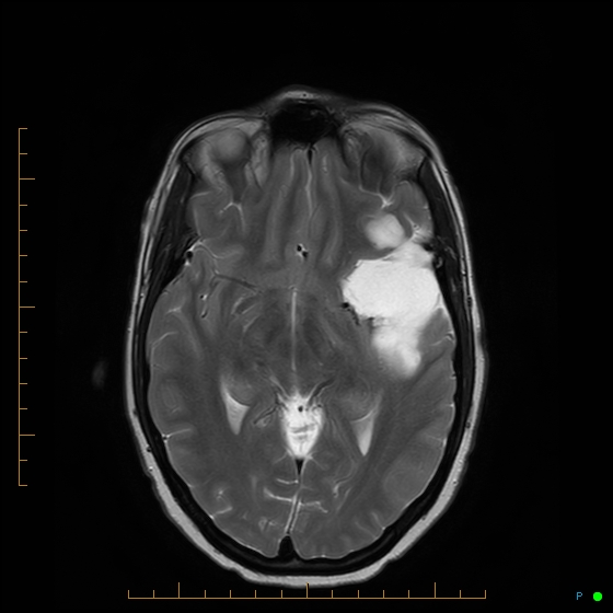
Large extra-axial, tumor-like lesion located in left frontotemporal area, expanding the Sylvian fissure with invasion of adjacent parenchyma at the frontal and temporal opercula with lobulated margins, with significant mass effect.
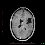
It demonstrates a very thin rim of enhancement post contrast infusion.The signal appearance of the lesion is similar to CSF and in diffusion weighted imaging demonstrates increased signal which is compatible with epidermoid growth.
The lesion demonstrates no evidence of hemorrhage or significant edema however, it is causing significant mass effect on the mesial temporal lobe, there is uncal herniation and incipient midline shift with mild compression of the left lateral ventricle.
Case Discussion
After a subtotal resection of the epidermoid tumor, histopathology confirmed the diagnosis of an epidermoid cyst.


 Unable to process the form. Check for errors and try again.