
Presentation
Recurrent headache, CT brain showed left cerebellopontine angle mass for MRI.
Patient Data


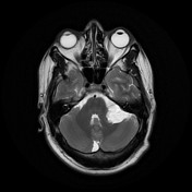
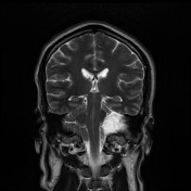

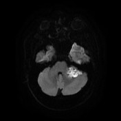
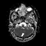
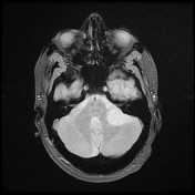
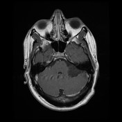

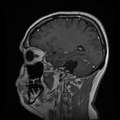
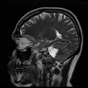
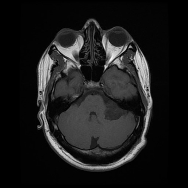
Left cerebellopontine angle, lobulated outline mass lesion shows the following characters; High signal on T2 and low signal on T1 (near isointense to CSF) it does not fully attenuate on FLAIR, instead of having a dirty, heterogeneous appearance. There is no contrast-enhancing component and it does not extend into the internal acoustic meatus (cranial nerves going through the lesion). On diffusion-weighted imaging (DWI) and (ADC) which demonstrates restricted diffusion. Exerting mild presser mass effect on the adjacent brain stem. The finding demonstrates typical appearances of left cerebellopontine angle epidermoid cyst.
Otherwise normal appearance of the cerebral hemispheres, corpus callosum, posterior fossa, with preserved gray/white matter differentiation.
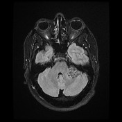


The FLAIR imaging is superior to conventional MR imaging in depicting intracranial epidermoid cysts, showing variable but satisfactory lesion contrast to both CSF and brain tissue.
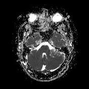
DW imaging reveals intracranial epidermoid tumors as hyperintense lesions relative to the brain and CSF and has the best conspicuity of the MR sequences. The hyperintensity of epidermoid tumors on DW imaging is not caused by only diffusion restriction in the lesions but by also the intrinsic T2 shine-through effect on the diffusion-weighted MR sequence.
Case Discussion
This is a typical case of an intracranial epidermoid cyst that matches with epidemiological characters and its most common presenting symptoms (headache).
Epidermoid cysts are well-demarcated, encapsulated lesions. The cysts may compress adjacent structures or they may be firmly anchored to them, sometimes encasing vessels and nerves.
Most of the epidermoid cysts are asymptomatic but depending on their location.
On imaging; the epidermoid cysts appear as well-demarcated lesions that resemble CSF and do not enhance with contrast agents. On diffusion-weighted images, they demonstrate restricted diffusion.
In this situation respect, MRI diffusion-weighted sequences.


 Unable to process the form. Check for errors and try again.