
Presentation
Non-Hodgkins lymphoma with CNS involvement subjected to bone marrow transplantation
Patient Data
Age: 60 years old
Gender: Male
From the case:
Intraosseous arachnoid granulations

Download
Info
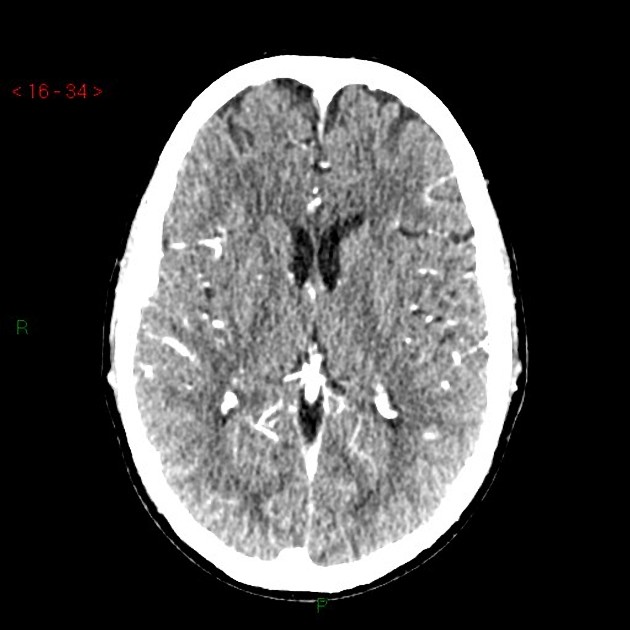
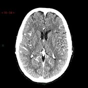
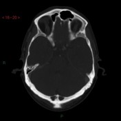
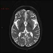
Occipital bone lytic areas are noted likely representing intraosseous arachnoid granulation
From the case:
Intraosseous arachnoid granulations
Download
Info
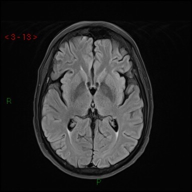
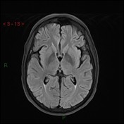
The described areas at the occipital bone in CT followed CSF signal intensity on MRI sequences.
Case Discussion
Prominent arachnoid granulations are an anatomical variant that could be mistaken as skull pathology.


 Unable to process the form. Check for errors and try again.