Note: This case has been tagged as "legacy" as it no longer meets image preparation and/or other case publication guidelines.
From the case:
Intraosseous hemangioma
{"study":{"id":11905,"case_slug":"intraosseous-haemangioma","modality":"X-ray","series":[{"id":571781,"content_type":"image/jpeg","frames":[{"id":571781,"width":379,"height":691,"current":true}],"annotations":[],"encodings":{"original_dicom":null,"video":null,"thumbnailed_files":[{"tiny":"a4edf38aeeda3dcce403edbc400d8c_tiny.jpg","jumbo":"a4edf38aeeda3dcce403edbc400d8c_jumbo.jpg","small":"a4edf38aeeda3dcce403edbc400d8c_small.jpg","thumb":"a4edf38aeeda3dcce403edbc400d8c_thumb.jpg","medium":"a4edf38aeeda3dcce403edbc400d8c_medium.jpg","gallery":"a4edf38aeeda3dcce403edbc400d8c_gallery.jpg","big_gallery":"a4edf38aeeda3dcce403edbc400d8c_big_gallery.jpg","original":"a4edf38aeeda3dcce403edbc400d8c.jpg"}]},"perspective":"Frontal","specifics":null},{"id":571792,"content_type":"image/jpeg","frames":[{"id":571792,"width":402,"height":689,"current":true}],"annotations":[],"encodings":{"original_dicom":null,"video":null,"thumbnailed_files":[{"tiny":"de0c702a2f0a308b94e9423eb69ab4_tiny.jpg","jumbo":"de0c702a2f0a308b94e9423eb69ab4_jumbo.jpg","small":"de0c702a2f0a308b94e9423eb69ab4_small.jpg","thumb":"de0c702a2f0a308b94e9423eb69ab4_thumb.jpg","medium":"de0c702a2f0a308b94e9423eb69ab4_medium.jpg","gallery":"de0c702a2f0a308b94e9423eb69ab4_gallery.jpg","big_gallery":"de0c702a2f0a308b94e9423eb69ab4_big_gallery.jpg","original":"de0c702a2f0a308b94e9423eb69ab4.jpg"}]},"perspective":"Oblique","specifics":null}],"caption":{"us":null,"gb":null},"findings":{"us":"\u003cp\u003eIntraosseous hemangioma of the distal phalanx. \u003c/p\u003e","gb":"\u003cp\u003eIntraosseous haemangioma of the distal phalanx. \u003c/p\u003e"},"contributor_login":"radswiki","case_id":11542,"case_key_image_id":null,"case_featured_at":null},"current_user":null,"access":{"can_edit":false,"can_download":true,"can_toggle_annotations":true,"can_feature":false,"can_pin":false}}
Intraosseous hemangioma of the distal phalanx.
Case Discussion
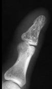
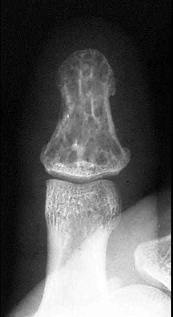
These images are from Dr. John Hunter's amazing MSK collection. Dr. John Hunter is a professor in the department of radiology (musculoskeletal section) at UC Davis School of Medicine.
This case was donated to Radiopaedia.org by Radswiki.net