
Presentation
A young female patient presented with left frontal supraorbital swelling for 2 years.
Patient Data
Age: 35 years
Gender: Female
From the case:
Intraosseous hemangioma - frontal bone


Download
Info
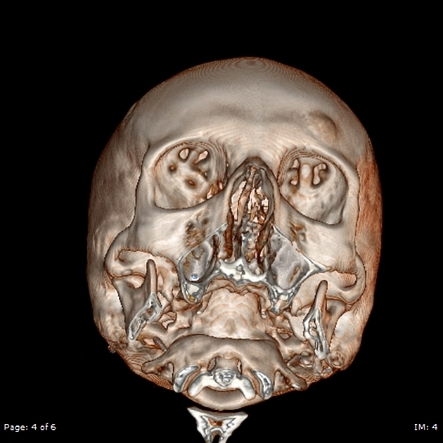
There is a well-defined lytic lesion involving the left frontal bone, measuring about 21 x 11 x 18 mm, it causes mild expansion of the left frontal bone. This lesion has coarsened trabeculation and sunburst pattern.
No intracranial extension of this lesion.
No soft tissue component or other malignant features.
Features are typical for left frontal intraosseous hemangioma.
Case Discussion
Intraosseous hemangiomas are more commonly seen in men and typically seen in the 4th to 5th decade of life.


 Unable to process the form. Check for errors and try again.