
Presentation
Gradually developed right-sided proptosis.
Patient Data
Age: 45 years
Gender: Female
From the case:
Intraosseous meningioma


Show annotations
Download
Info
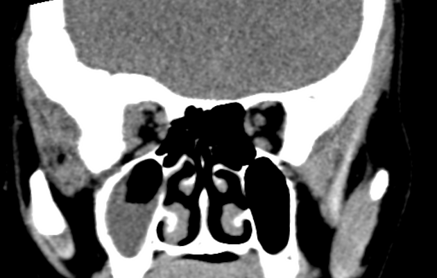
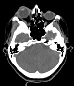
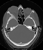
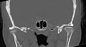
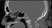
There is bony expansion, remodelling, and hyperostosis with surface irregularity involving the greater wing of the right sphenoid bone, squamous part of the right temporal bone, right frontal bone, and superolateral orbital wall causing right exophthalmos, without obvious soft tissue component.
Eye globes and optic nerves are grossly unremarkable.
Partial opacification of the right maxillary sinus is seen.
Case Discussion
CT findings are most likely suggestive of sphenoid wing intraosseous meningioma enplaque as described above.
Co-contributor: Dr. Anwar-ul-Haq Zadran.


 Unable to process the form. Check for errors and try again.