
Presentation
Incidental finding in a female patient with pelvic pain for about ten days.
Patient Data
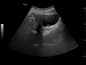
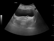
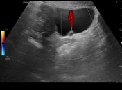
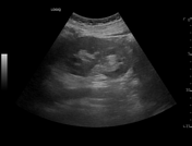
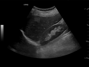
Transabdominal color doppler US image shows anechoic cystic dilatation of the distal left ureter and the presence of a ureteric jet (ureteric jet angle of approximately 90°) indicates the absence of complete obstruction. Both kidneys are normal. In segment 6 of the liver, there is a 2.5 cm hemangioma.
Radiographer: TSRM Nunzio Bianco
Case Discussion
A ureterocele is visible as a cystic lesion in the bladder 2. Diagnosis of ureterocele is helpful because it can cause obstruction, urinary tract infection, and hydronephrosis 1. Treating ureterocele prevents complications that can lead to kidney failure 1. Ureteric jets are normally symmetrical, valid (if they cross the midline) and are not synchronous. The identification of the ureteric jet excludes the presence of complete obstruction.


 Unable to process the form. Check for errors and try again.