
Presentation
Screening mammogram
Patient Data

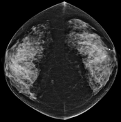
There is extremely dense fibroglandular tissue (BI-RADS D).
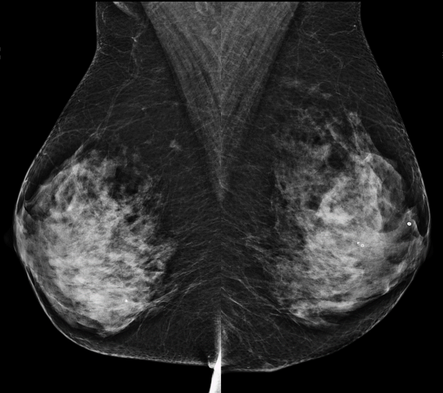
There is a small ill-defined mass in the right upper inner quadrant in the retroglandular space, anterior to the pectoral muscle. On the CC view, the mass is seen in the medial breast and, therefore, falls in two of the forbidden zones on mammography.
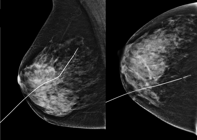
This is best regarded as an M4 lesion and should be recalled for assessment.
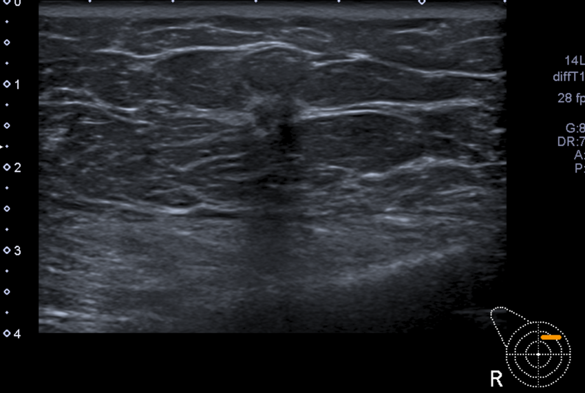
An irregular mass is seen in the right upper inner quadrant. This causes posterior acoustic shadowing and interruption of the fat planes (U5).
The axillary lymph nodes were normal, with a thin cortex and preserved hilum (not shown).
Case Discussion
There are four forbidden zones on mammography:
anterior to the pectoral muscle (milky way)
behind the nipple
retroglandular space
medial breast
Lesions that fall within these regions should be regarded as suspicious for cancer as there is less glandular breast tissue in these locations. This is particularly so if the lesion is new in comparison to the previous screening mammogram.
The lesion was confirmed to be a grade 2 invasive ductal carcinoma at surgical excision.


 Unable to process the form. Check for errors and try again.