
Presentation
Presents with intermittent complaints of pain in epigastric region after meals.
Patient Data


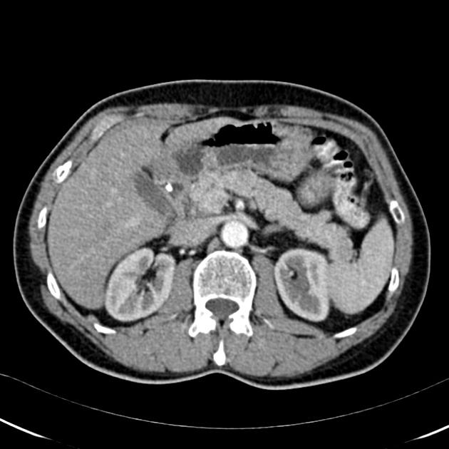
In the late arterial phase, a solid lesion is noted at the pancreatic neck which is homogeneously iso- to mildly hyperdense compared to the pancreatic parenchyma.
In the venous phase, the lesion is also seen as a well-circumscribed homogeneously hyperdense (hypervascular) mass. It does not cause pancreatic duct obstruction or dilatation.
Case Discussion
Islet-cell tumor or neuroendocrine tumor of the pancreas.
The typical appearance of these tumors is well demonstrated in the venous phase. This hypervascular lesion demonstrates late retention of contrast and typically has a soft texture, therefore not causing pancreatic duct obstruction or dilatation.
Pancreatic islet-cell tumors account for about 1-5% of all pancreatic neoplasms. They represent an important subset of neoplasms due to their substantially improved prognosis compared to pancreatic adenocarcinoma.
PET-CT findings were negative in this patient.
Endoscopic ultrasound demonstrated a well-circumscribed pancreatic tumor of 11 x 14 mm, FNA cytology was performed and confirmed the diagnosis.


 Unable to process the form. Check for errors and try again.