
Note: This case has been tagged as "legacy" as it no longer meets image preparation and/or other case publication guidelines.
From the case:
Jugulotympanic paraganglioma
Download
Info
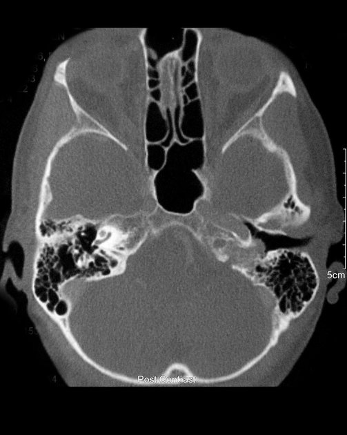
A single axial CT bone image through the temporal bones demonstrates a mass arising from the jugular bulb region and extending into the middle ear over the cochlear promontory. The bony margins are irregular and spiculated.
From the case:
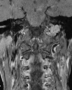
Jugulotympanic paraganglioma

Download
Info

Single axial and coronal images demonstrate a lobulated enhancing (confirmed on T1C- not shown) mass in the left temporal bone centred on the jugular foramen.
From the case:
Jugulotympanic paraganglioma
Download
Info
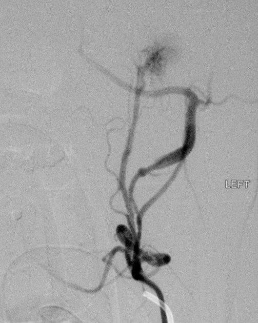
Left external carotid artery injection demonstrates a high vascular blush.
Case Discussion
Features on CT, MRI and DSA are consistent are those of a jugulotympanic paraganglioma. Unfortunately, follow-up and/or histological confirmation are not available.


 Unable to process the form. Check for errors and try again.