
Presentation
Bilateral hip region and left knee joint pain for the last 6 months.
Patient Data
Age: 10 years
Gender: Female


Show annotations
Download
Info
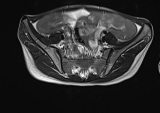
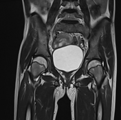
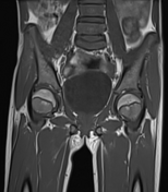
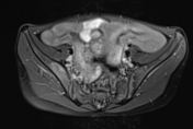
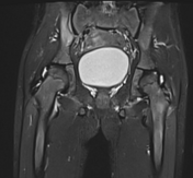
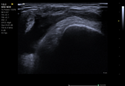
There is edema in one of the muscles of the quadriceps femoris near its origin in the left proximal femur region, likely enthesitis.
Show annotations
Download
Info

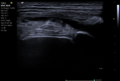
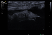
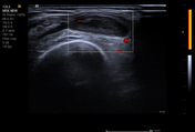
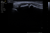
The ultrasound of the left knee joint reveals mild joint effusion and mild diffuse synovial thickening with minimal internal vascularity of power Doppler, suggesting active synovitis.
Case Discussion
The rheumatoid factor and ENA profile of the patient were negative, and HLA-B27 was positive. The ESR and CRP were high.
The patient had significant improvement in her symptoms after steroid therapy.
Co-author: Dr. Vivek Ranjan (pediatrician).


 Unable to process the form. Check for errors and try again.