
Presentation
History withheld.
Patient Data
Note: This case has been tagged as "legacy" as it no longer meets image preparation and/or other case publication guidelines.
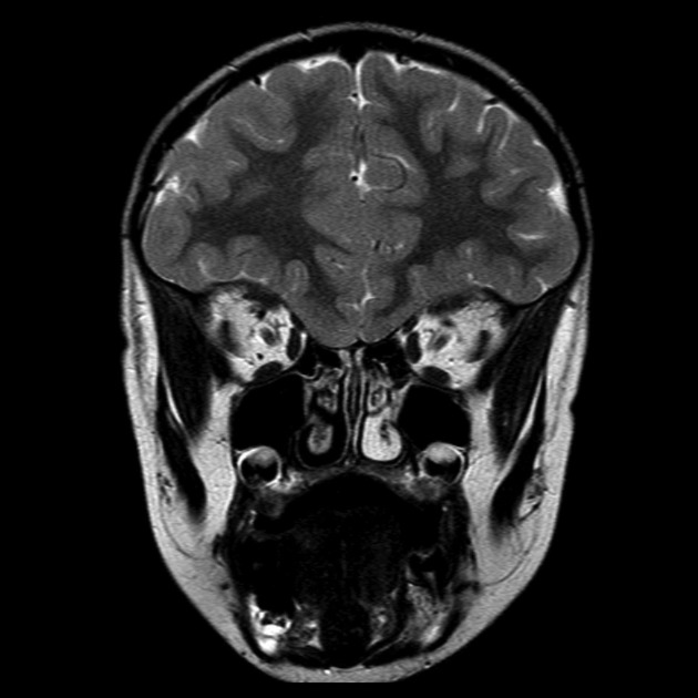
Single coronal T2 image through the frontal lobes demonstrates abnormal anatomy with absence of the olfactory bulbs and olfactory sulcus. The gyrus rectus and medial orbital gyrus form a single gyrus.
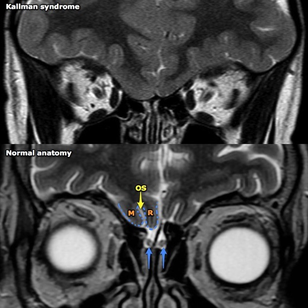
It is easiest to appreciate the anatomical anomalies present in Kallman syndrome by comparing it to a normal patient. The normal anatomy of the region consists of the olfactory bulbs (blue arrows) located in the olfactory grooves of the anterior cranial fossa. The inferior surface of the frontal lobes usually consists of gyrus rectus (aka straight gyrus) (R) separated from the medial orbital gyrus (M) by the olfactory sulcus (yellow arrow). These are absent in Kallman syndrome.
Case Discussion
Kallmann syndrome is rare, but has characteristic imaging findings which should be sought in patients with delayed onset of puberty/hypogonadism.


 Unable to process the form. Check for errors and try again.