
Presentation
Radial-sided right wrist pain.
Patient Data
Age: 25 years
Gender: Male
From the case:
Kienbock disease


Download
Info

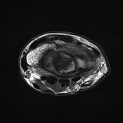
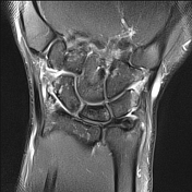
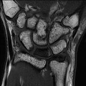
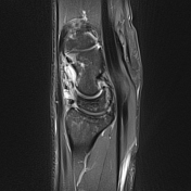
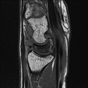
Alignment is within normal limits. No fracture. No focal osseous lesion. Radiocarpal effusion with synovial thickening. No distal radioulnar joint effusion. Distinct low T1 signal of the lunate with mild bone marrow edema; no collapse. Punctate periarticular high signal diffusely through the wrist. Capsular edema including the extrinsic ligaments.
Case Discussion
Signal change is typical of Kienbock disease (lunate osteonecrosis) although without collapse or fragmentation. Presumably, the joint effusion is reactive with synovial thickening likely represents synovitis. Diffuse punctate periarticular high signal may be due to disuse osteopenia.


 Unable to process the form. Check for errors and try again.