
Presentation
Gradual onset of left side painless proptosis was observed.
Patient Data
Age: 50 years
Gender: Female
From the case:
Lacrimal gland lymphoma


Download
Info

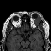
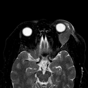
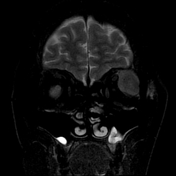
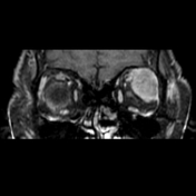
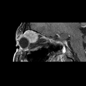
A well circumscribed left orbital extra conal mass lesion occupying the lacrimal fossa and compressing the left lateral rectus inferomedially, the superior rectus medially and the globe anteriorly. The lesion returns intermediate signal on T1, T2 and STIR with moderate enhancement on post contrast study.
No evidence of fat stranding, orbital or bony invasion.
Case Discussion
Histopathological specimen from the excised mass revealed MALT lymphoma. The imaging features are typical for lymphoma as the high cellularity of the tumors renders them of intermediate or low signal on T2 WI. Unfortunately DWI wasn't available in this study although it would be of great help.


 Unable to process the form. Check for errors and try again.