
Presentation
Generalized lethargy. Previous stroke 2 month earlier.
Patient Data
Age: 80 years
Gender: Female
From the case:
Laminar cortical necrosis

Download
Info

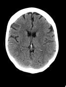
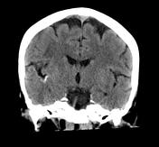
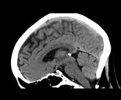
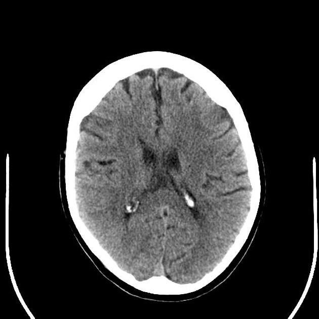
Gyriform high density in the right parietal and temporal lobes, with underlying white matter hypodensity, correlate to the regions of ischemia seen on the previous MRI.
Download
Info

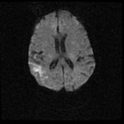
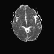
Several areas of restricted diffusion in the right frontal lobe, right insula, right parietal lobe are in keeping with acute infarctions in the right MCA territory. The largest focal areas of diffusion restriction are in the right parietal lobe, however there are punctate foci elsewhere in the MCA territory.
Download
Info
There is subtle hypoattenuation and loss of grey-white matter differentiation in the posterior right insular cortex and superior temporal lobe.


 Unable to process the form. Check for errors and try again.