
Presentation
Routine head MRI for non-specific headaches.
Patient Data


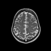
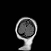
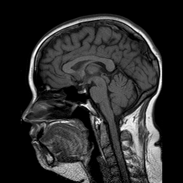
Round T2-iso-hyperintense with partial suppression on FLAIR and T1-hypointense mass is located posterior to precuneus sagittally, splitting the superior sagittal sinus, well seen on sagittal MRV.
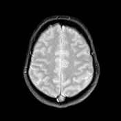
There are some small round T2 and FLAIR hyperintense foci within frontal and parietal lobes bilaterally, depicting small vessel ischemia.
There is a small blooming artifact within pineal gland: calcium deposition.
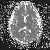
No abnormal signal on DWI and ADC.
The remainder brain seems unremarkable.
Case Discussion
This is nice case of normal anatomical structure with its increased size and uncommon sagittal localization within the superior sagittal sinus. It can be easy mistaken for dural venous thrombosis, for the avoidance of it, an MRV study can be performed with 15-10-5 cm/s velocity settings.


 Unable to process the form. Check for errors and try again.