
Presentation
History of dermatomyositis presents with a severe headache. Left pyramidal syndrome and fever are documented on admission.
Patient Data
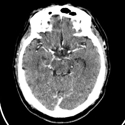
Contrast CT shows a poorly defined hypodense lesion at the level of the right thalamus that results in compression of adjacent structures with slight perilesional edema and does not show enhancement.
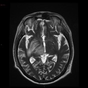

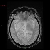
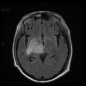
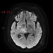
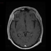

MRI shows a rounded intra-axial lesion, hypointense in T1 with no evidence of blood, corroborated on echo gradient. FLAIR sequence demonstrates edema and at the ipsilateral mesencephalic level. In DWI and ADC there is a restriction in the central portion of the lesion and with the application of contrast medium a fine annular enhancement is observed.
Case Discussion
Blood cultures confirmed infection with Listeria. These features are consistent with late cerebritis early abscess formation with central liquefaction and surrounding enhancing capsule formation.


 Unable to process the form. Check for errors and try again.