
Presentation
MRI was originally performed for medial knee pain.
Patient Data
Age: 30 years
Gender: Female
From the case:
Lateral discoid meniscus


Download
Info
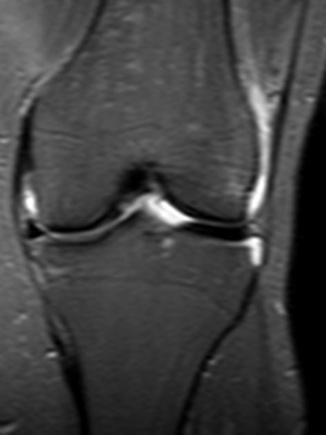
Discoid lateral meniscus.
Horizontal high T2 intrasubstance signal at the posterior horn of medial meniscus not reaching the articular surface, reflecting grade II meniscal degeneration.
Case Discussion
In this case, meniscal degeneration was more advanced in the medial posterior horn than in the rather incidentally found lateral discoid meniscus.


 Unable to process the form. Check for errors and try again.