
Lateral patellar dislocation with lipohemarthrosis on ultrasound
Presentation
Awkward fall off bicycle on to the left knee. The knee is swollen and he is unable to straight leg raise. ?Patella tendon injury
Patient Data
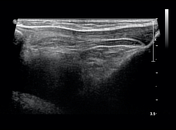

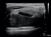
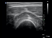

The patella and quadricep tendons are intact. Evidence of a large knee joint effusion with layering fluid of different echogenicities in the suprapatellar pouch. The most superficial of these fluids is high echogenicity, consistent with fat. Findings represent a lipohemarthrosis.
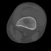
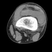
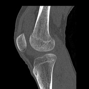
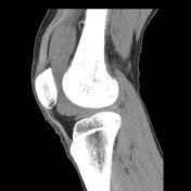
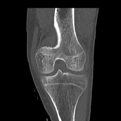
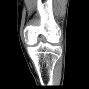

Large lipohemarthrosis with minimally impacted fractures of the medial aspect of the medial patellar facet, and the lateral aspect of the lateral femoral condyle. Adjacent to the medial patella are tiny fracture fragments, with additional tiny fragments anterior to the tibial plateau close to the midline, and adjacent to the posterior third of the lateral meniscus. Findings are likely consistent with a recent lateral patella dislocation.
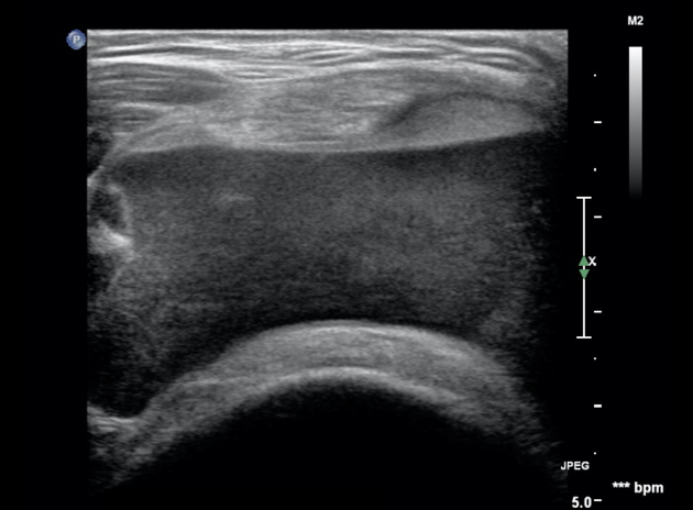
Annotated study which highlights the fat-fluid level within the suprapatellar portion of the knee joint.
Case Discussion
Injury to the extensor mechanism; the quadriceps tendon, patella, and patella tendon, or a large knee joint effusion, are potential causes that prevent the perform a straight leg raise (SLR). Clinical examination can be equivocal and the lack of patella displacement in partial thickness injuries necessitates the need for further imaging.
A lipohemarthrosis signifies an intra-articular injury, and whilst many will be accustomed to identifying a lipohemarthrosis on a horizontal beam lateral knee radiograph, it can be overlooked on ultrasound.
Similarly, many will be used to identifying the different bone marrow contusion patterns in knee injuries on MRI 1, and less so on CT. Having a high index of suspicion and a structured approach with CT interpretation can identify the injuries sustained in a recent lateral patella dislocation.


 Unable to process the form. Check for errors and try again.