Presentation
Suspicious of von Recklinghausen disease on CT scan.
Patient Data


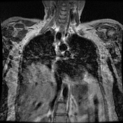

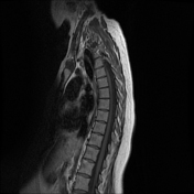

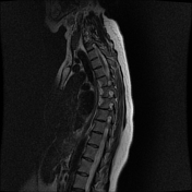

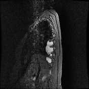

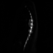
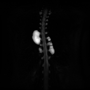
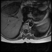

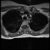


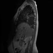

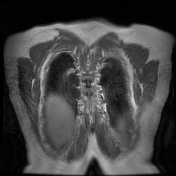

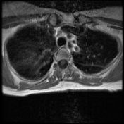

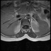

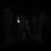

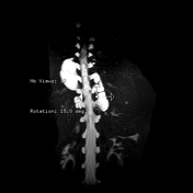

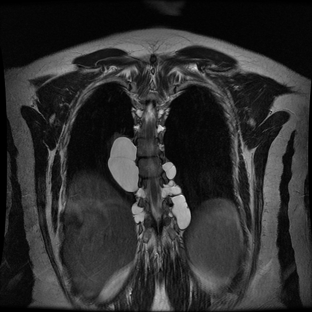
Bilateral lateral meningoceles are seen within neural foramina at visualized thoracic levels extending into extra-foraminal spaces at multiple levels and causing mass effects upon adjacent parts of the lungs.
All these meningoceles appear isointense to CSF in all sequences with no evidence of abnormal enhancement after contrast injection.
Kyphosis is seen in the thoracic spine.
T1-2 to T12-L1 levels are essentially unremarkable.
The spinal cord appears within normal limits in shape, size, and signal intensity, it ends at T12.
CSF flow artifacts were noted in the T2WI images as decreased signal intensities especially behind the cord.
Case Discussion
Bilateral lateral meningoceles are seen within neural foramina at visualized thoracic levels.
This disorder manifests itself with formations of cysts at different levels of the central nervous system along with meningeal diverticula protruding through the intervertebral spaces and filled by cerebrospinal fluid (CSF).


 Unable to process the form. Check for errors and try again.