
Presentation
Persistent atrial fibrillation
Patient Data
Age: 40 years
From the case:
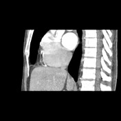
Left atrial diverticulum


Download
Info
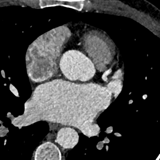
Small left atrial diverticulum extending from superior wall.
CT coronary angiography is normal with no evidence of coronary atherosclerosis. Normal coronary arteries.
Case Discussion
A left atrial diverticulum is a normal variant. It is important to be aware of to avoid interpreting it as pathology.


 Unable to process the form. Check for errors and try again.