
Presentation
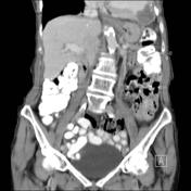
Incidental finding on CT study of the abdomen.
Patient Data
Age: 85 years
Gender: Female


Download
Info
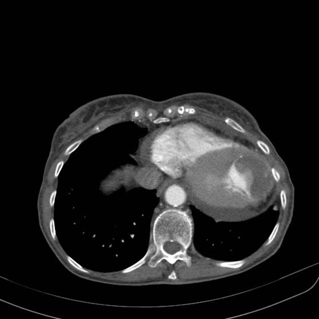
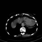
Aneurysmal dilation of the cardiac apex partially filled by a thrombus. The wall of the aneurysm is thinner than the wall of the rest of the left ventricle.
Case Discussion
A left ventricular aneurysm refers to a discrete, dyskinetic area of the left ventricular wall with a broad neck (as opposed to left ventricular pseudoaneurysms, thus often termed true aneurysms)


 Unable to process the form. Check for errors and try again.