
Presentation
Follow up post inferior STEMI
Patient Data
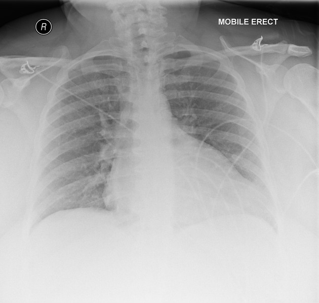
Lungs and pleural spaces are clear. No focal consolidation or collapse.
The cardiac silhouette is enlarged even allowing for AP projection with a focal bulge at the apex.


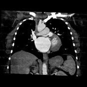
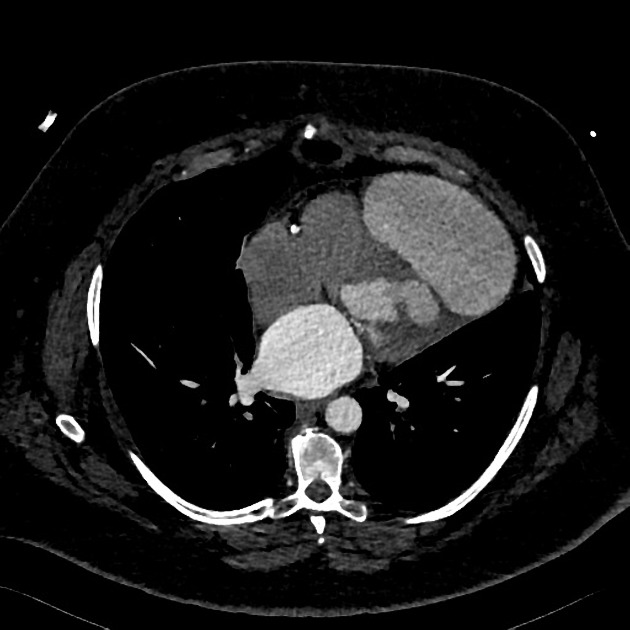
Large (9.6 x 9.1 cm) aneurysm extends from an 11 mm defect in the posterior aspect of the aneurysmal inferior left ventricular wall with adjacent fatty metaplasia. The aneurysm displaces the left ventricle superiorly and there is surrounding stranding in the epicardial fat. More superiorly in the anterior wall is a 7 mm region of wall thinning, possibly the site of further aneurysm development.
Mixed calcified and non-calcified atheroma is present throughout the coronary arteries.
Case Discussion
The patient had a previous myocardial infarct.


 Unable to process the form. Check for errors and try again.