
Presentation
Abdominal pain and fever.
Patient Data
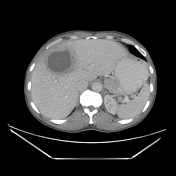

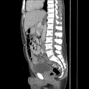
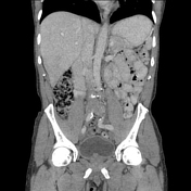
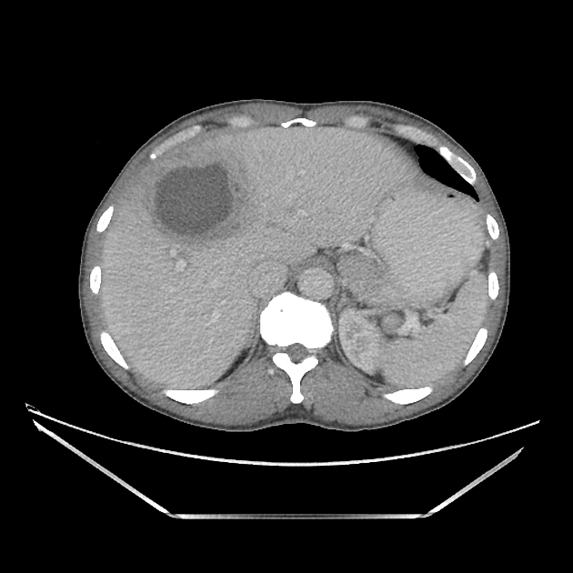
Large liver mass in the anterior right hepatic lobe, involving segments 8, 5, and 4, with central low-attenuation, peripheral irregular rind-like enhancement, and surrounding hepatic parenchymal edema. There is thrombosis of the middle hepatic vein leading into and coursing along the posterior inferior margin of this mass, with a small amount of clot leading into the inferior cavoatrial junction. There is a similar mass in hepatic segment 7, slightly smaller. Some generalized heterogeneous/diminished perfusion in the segment supplied by the thrombosed middle hepatic vein. There is focal fat along the falciform ligament. No other intra-abdominal abnormalities.

Ultrasound images demonstrate the correlate for the mass, with similar hypoechoic center, peripheral rind, and parenchymal edema. These images were obtained as part of a guided percutaneous drainage of the liver abscess.
Case Discussion
Liver abscesses can be unexpected and challenging to diagnose and have a variety of appearances. Though in this setting, the relatively low attenuation center, peripheral enhancement, and inflammatory surrounding reaction that we typically associate with abscess elsewhere in the abdomen/pelvis are present, which helps to increase confidence in this diagnosis. The thrombosis of the middle hepatic vein with a small amount of clot leading into the cavoatrial junction is an important finding not to overlook!


 Unable to process the form. Check for errors and try again.