
Presentation
6 months of slow growing mass above right ankle.
Patient Data
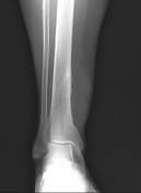
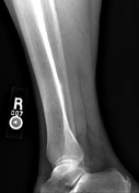
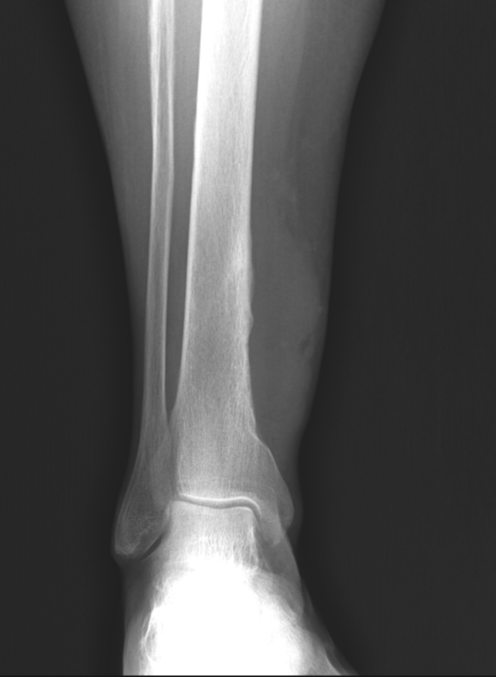
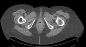
On the frontal view, there is a large soft tissue mass along the distal medial aspect of the lower leg with a periosteal reaction involving the medial aspect of the distal right tibia.
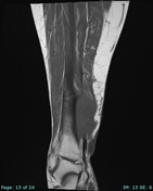

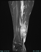
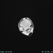
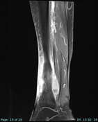

Large soft tissue mass abutting the medial cortex of the distal tibia. Mass is T1 isointense to muscle and T2 hyperintense. Mass avidly enhances following contrast administration.
The tibial cortex appears intact. Abnormal edema and enhancement is present in the underlying marrow.
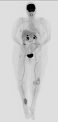
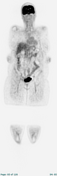
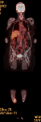

Right lower leg soft tissue mass is moderately FDG avid with SUV max of 7.5 (reference liver SUV max 4.2).
No enlarged or FDG avid inguinal lymph nodes.
No evidence of distal metastases.
Unrelated inflammatory FDG uptake about the left knee.
FINAL DIAGNOSIS: Leiomyosarcoma, high grade.
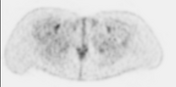
Given the morphologic features of a smooth muscle neoplasm on H&E in conjunction with positivity for smooth muscle markers (strongly positive for caldesmon and muscle specific actin, and weakly positive for smooth muscle actin), a diagnosis of leiomyosarcoma is supported. The presence of necrosis and nuclear pleomorphism are consistent with at least grade 2 by FNCLCC criteria.
Case Discussion
FDG PET-CT can be useful in the setting of soft-tissue sarcomas, such as leiomyosarcoma, for biopsy guidance, grading, staging, response assessment and surveillance.


 Unable to process the form. Check for errors and try again.