
Presentation
Mechanical fall with "popping" sound in ankle. Right medial ankle tenderness. Tenderness in right fibula head.
Patient Data

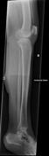
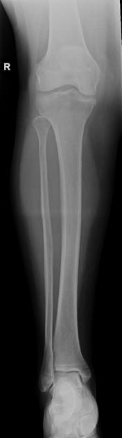
Undisplaced spiral fracture through the proximal fibula. Undisplaced transverse fracture through the medial malleolus. Distal tibiotalar joint appears intact.
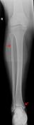
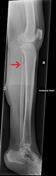
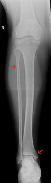
In addition to the fractures, there is a "stitching line" (indicated by arrows). This causes an artifact in which there appears to be a cortical defect in the fibula, which could be mistaken for a fracture line. There is also linear increased density in the soft tissues adjacent, also an artifact.
Case Discussion
The combination of a proximal-third fibula fracture and medial malleolus fracture has the eponymous name of a Maisonneuve fracture.
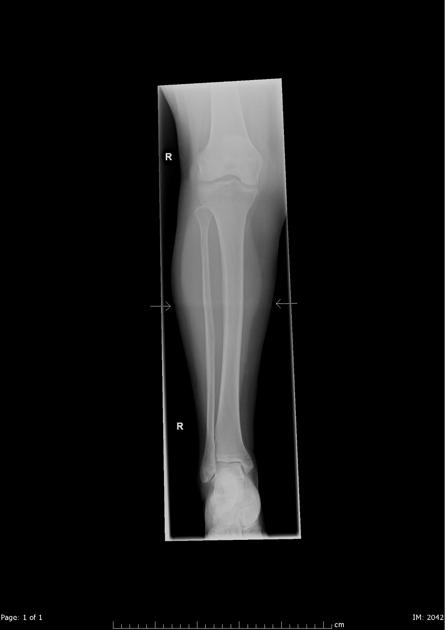
This case also demonstrates the importance of two orthogonal views to detect and/or confirm the location of pathology. When reading CR or DR images it is important to be aware that some long-leg views are automatically stitched together and if this is imperfect, artifacts that mimic pathology may occur.
The patient required operative management with two medial malleolus screws and a diastasis screw.


 Unable to process the form. Check for errors and try again.