
Presentation
History of dementia, presenting with a large mass and a bleeding wound on the right first toe. This has been growing/worsening over the past year. The right first toenail was removed a year ago due to a reported ingrown toenail.
Patient Data
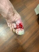


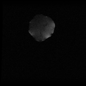
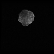
Pictures of the patient's right foot show a large protuberant mass at the distal aspect of the first toe. The proximal aspect of the toe also appears irregular with areas of bleeding.

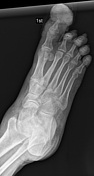
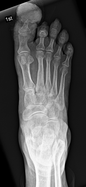
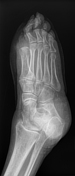
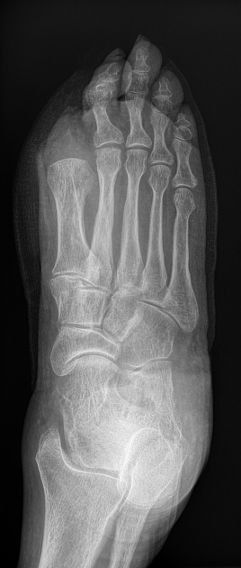
Initial radiographs of the right foot show a mass at the distal first toe with overlying dressing material. Mild truncation of the hallux distal phalanx tuft is noted, likely due to chronic erosion.
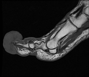

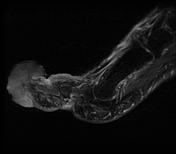
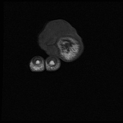
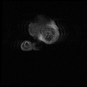
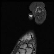
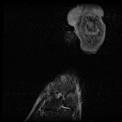


MRI of the right forefoot without and with IV contrast demonstrates a large exophytic mass at the distal aspect of the first toe in the cutaneous/subcutaneous tissues. The mass demonstrates mild T1 hyperintensity relative to skeletal muscle, mild T2 hyperintensity, and predominantly peripheral enhancement and septal enhancement. There is skin thickening around the hallux with circumferential enhancement. Mild T2 hyperintensity in the hallux distal phalanx without corresponding T1 hypointensity likely represents reactive marrow oedema. Mild truncation of the hallux distal phalanx tuft is noted, likely due to chronic erosion. Motion artifact degrades several sequences, greatest on the axial T2 fat sat and coronal STIR sequences.

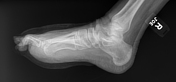
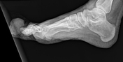
Subsequent radiographs of the right foot show amputation of the hallux at the level of the metatarsophalangeal joint. The hallux sesamoids were also removed. There is soft tissue swelling at the amputation site.
Case Discussion
Pathology from the hallux amputation showed malignant melanoma, nodular type. No definite perineural or lymphovascular invasion was identified. No metastases were found.
Malignant melanoma, a potentially deadly form of skin cancer originating from melanocytes (the pigment-producing cells), can manifest on any part of the body. Malignant melanoma of the hallux (great toe) may initially be misdiagnosed as infection or haematoma 1.
CT, MRI, and PET are valuable tools for comprehensively evaluating malignant skin tumours, including melanoma. These advanced imaging techniques are instrumental in determining the extent of the primary tumour, assessing the involvement of regional lymph nodes, and identifying any potential distant metastatic spread throughout the body 2.
The definitive diagnosis of malignant melanoma of the toe is established through a biopsy. Once the diagnosis is confirmed, the treatment approach involves a wide radical surgical excision to remove the tumour and a surrounding margin of healthy tissue 3.


 Unable to process the form. Check for errors and try again.