
Presentation
Recent severe left otitis externa, swelling extending to the left face. Now septic. History of type 2 diabetes and smoking.
Patient Data


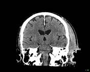
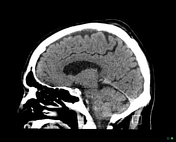
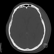
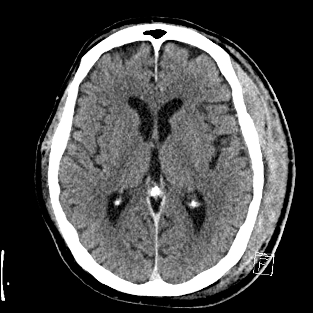
No intra/extra-axial collections. No midline shift or herniation. Grey-white matter differentiation is preserved. No enhancing intracranial lesions. The ventricles and CSF spaces are normal. No features of thrombosis of the major dural venous sinuses. The orbits appear normal. Paranasal sinuses are well-aerated.
Soft tissue thickening along the left external auditory canal. Extensive skin thickening and subcutaneous inflammatory change in the pre and post auricular regions. Heterogeneously enhancing soft tissue thickening extending superiorly along the temporalis and subgaleal space of the temporal bone. No appreciable focal hypodense collection within the region.
Partial opacification of the left mastoid air cells. No appreciable bony erosions of the external acoustic canal. The middle ear structures appear grossly intact.
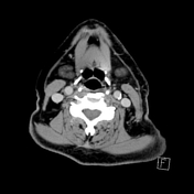
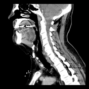
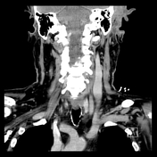

Fat inflammatory stranding within the left parotid space with early extension to the parapharyngeal space and nasopharynx. No appreciable collection within this region.
Multiple prominent pre and post auricular lymph nodes, likely reactive. The salivary glands and thyroid display normal enhancement. No other acute abnormality.
Impression
Left necrotizing otitis externa with inflammatory change involving the parotid, masticator space and early extension to the left parapharyngeal space. Superior extensive inflammatory phlegmon along the temporalis and temporal subgaleal region. No appreciable collection at present. Evidence of left mastoiditis with no intracranial extension.
Case Discussion
The patient recovered slowly following long-term IV antibiotics and did not require surgery.


 Unable to process the form. Check for errors and try again.