
Presentation
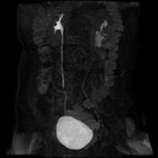
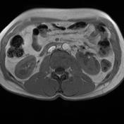
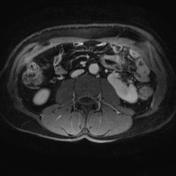
Left varicocele. Suspicious left kidney on ultrasound.
Patient Data
Age: 35 years
Gender: Male
From the case:
Malrotated kidney


Download
Info

Left malrotated kidney, where the hilum faces anteriorly.


 Unable to process the form. Check for errors and try again.