
Presentation
MRI brain for surgical planning of left posterior fossa meningioma.
Patient Data
Age: 60 years
Note: This case has been tagged as "legacy" as it no longer meets image preparation and/or other case publication guidelines.
From the case:
Mastoid segment facial nerve schwannoma
Download
Info

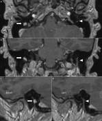
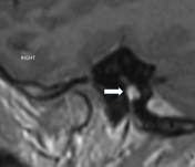
MRI brain gadolinium enhanced T1 weighted images demonstrate an enhancing nodule in the mid segment of the right facial nerve (arrow). The normal mastoid segment of the left facial nerve is also shown (arrow head). This is an incidental finding and the evaluated left posterior meningioma is not shown.
Case Discussion
Facial nerve schwannoma is a rare benign tumor. As schwannomas elsewhere it originates from the surface of the nerve, and displace and splay the nerve fibers over their eccentric growth.


 Unable to process the form. Check for errors and try again.