
Presentation
Irregular menstrual period and lower abdominal pain.
Patient Data

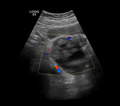
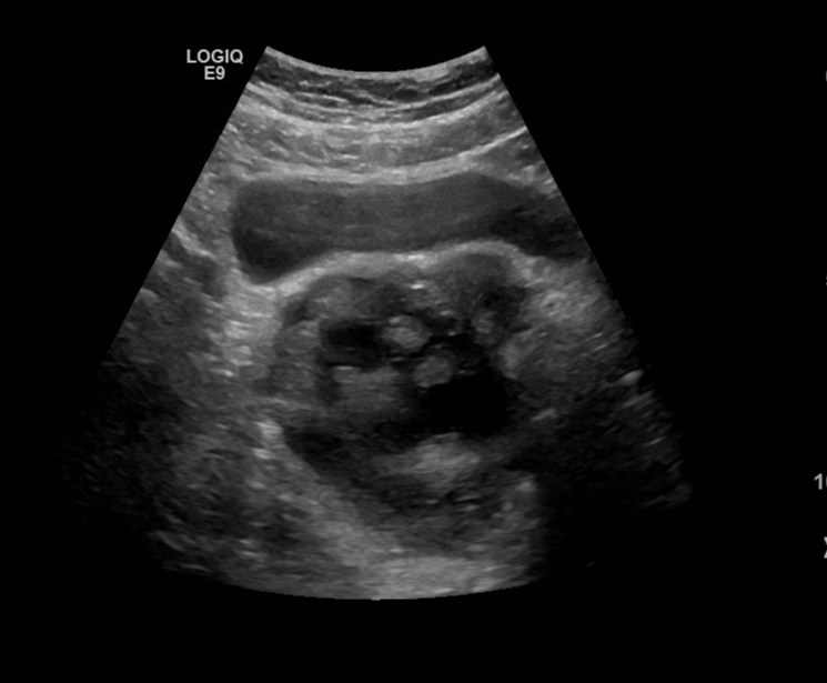
There is a right adnexal cystic lesion that shows multiple echogenic floating balls.
There was no vascularization within the balls.
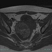


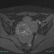

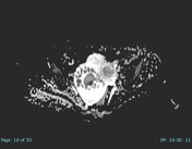
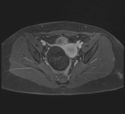

There is a cystic lesion arising from the right ovary showing multiple internal rounded lesions which appear of higher signal than the fluid on T1W and lower signal than fluid on T2 W images giving the appearance of floating balls sign, with evidence of restricted diffusion, and fat components, without evidence of enhancing solid component.
It measures 7 x 5.4 x 6.4 cm. It pushes the uterus and sigmoid to the left side.
Features are suggestive of right ovarian mature cystic teratoma.
Thin-walled fluid intensity cyst arising from left ovary measuring 4.7 x 3.6 x 5 cm.
Minimal pelvic free fluid.
Normal thickness of the endometrium.
No uterine masses.
Case Discussion
In rare cases, the presentation of MDCT is atypical, which can be a diagnostic challenge for radiologists.
Multiple small floating spheres within a large cyst, as observed in the case presented here, is one of those rare presentations, known as the “floating balls” presentation but it is pathognomonic for ovarian mature cystic teratoma.


 Unable to process the form. Check for errors and try again.