
Presentation
Mainly right-sided deficits, such as hemiparesis, decreased sensation, and facial droop.
Patient Data


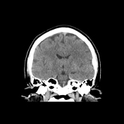
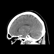
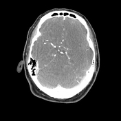
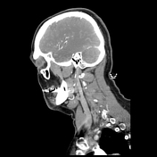
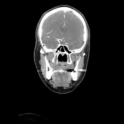
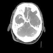
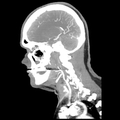
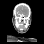
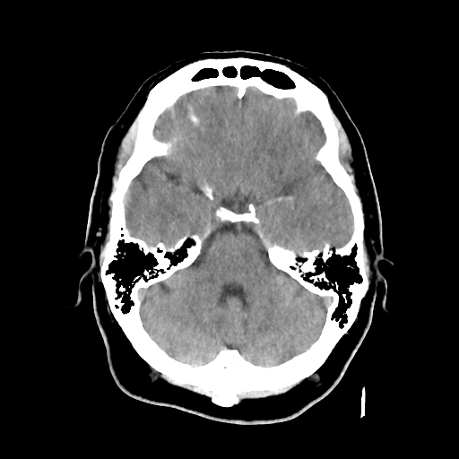
There is loss of gray-white matter differentiation involving the left frontal lobe, operculum, insular cortex and basal ganglia indicative of an acute infarct. There is a hyperdense left middle cerebral artery involving the M1 and M2 segments.
There is left internal carotid artery occlusion with the partial opacification of the petrous segment. There is partial reconstitution of the internal carotid artery mid cavernous segment. There is subsequent occlusion of the left ICA terminus with thrombus extending into the proximal left ACA
A1 segment and the left MCA M1 and M2 segments. There is minimal opacification of the distal left MCA M2 and M3 branches in the left parietal region.
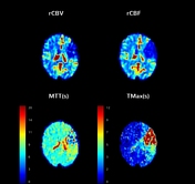
On CT perfusion, there is a left frontal lobe core infarct with a larger left cerebral hemisphere area of ischemia.
Case Discussion
This is a case of the MCA dot sign, which indicates a visible thrombus in the MCA. This finding corroborated with the CTA and CT perfusion.


 Unable to process the form. Check for errors and try again.