
Presentation
Hemiplegia.
Patient Data
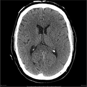


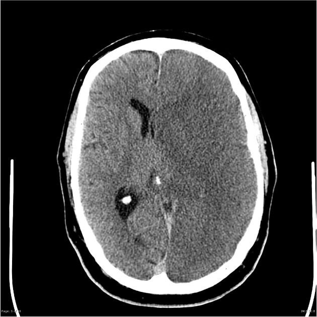
Non-contrast CT shows no acute hemorrhage. Loss of grey-white differentiation at the caudate head and insula ribbon. Hyperdense left M1 segment.
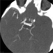
CTA shows a filling defect in the left M1 segment in keeping with acute thrombus.
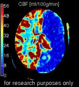
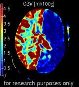

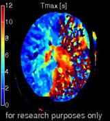
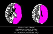


There is a large region of reduced CBV, CBF and time to peak perfusion affecting the whole left cerebral hemisphere. This is in keeping with a large left MCA infarct. A very small region of mismatch reduced time to peak perfusion is suggestive of only a tiny penumbra.
Conclusion
Findings are in keeping with a large left hemispheric infarct.
Expected evolution of known left MCA territory infarct with left cerebral hemisphere loss of grey-weight differentiation with edema and positive mass effect. Hyperdense left MCA again demonstrated. No hemorrhagic transformation.
Case Discussion
This case demonstrates acute changes with acute MCA territory infarct:
- hyperdense MCA sign
- loss of grey-white differentiation, seen earliest in the insular cortex and basal ganglia
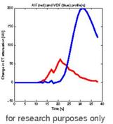
Perfusion imaging is more commonly performed to calculate the presence and size of the ischemic penumbra (i.e. cerebral parenchyma at risk of infarction but not yet infarcted) through which various interventions (e.g. thrombolysis, clot retrieval, etc) can be considered for treatment with the aim of improved outcomes.


 Unable to process the form. Check for errors and try again.