
Presentation
Sudden onset left weakness 40 minutes ago.
Patient Data


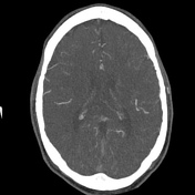
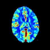
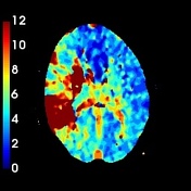
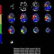
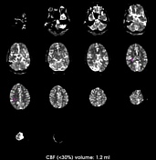
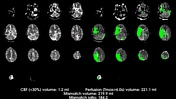
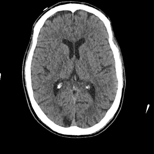
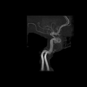
Right distal ICA and M1 segment MCA thrombosis seen on CTA without obvious features of infarction on the non-contrast CT but with evidence of a large area of abnormal perfusion (Tmax>6s, estimated volume 221ml) and small infarct core (CBF<30%, estimated volume 1.2ml) giving an estimated ischemic penumbra of 220ml. The patient proceeded to the angiography suit for clot retrieval.

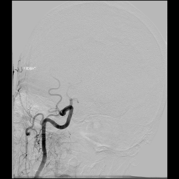
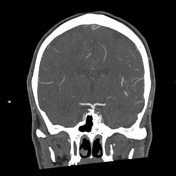
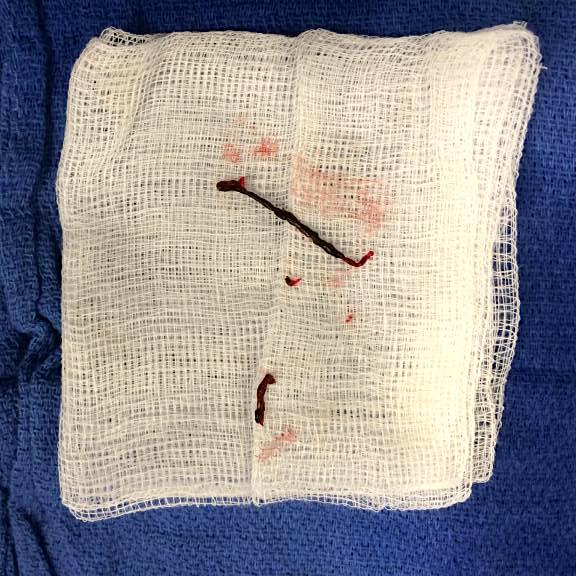
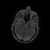
Prior to clot retrieval right ICA injection revealed near complete occlusion of the right distal ICA with no flow into the right MCA. After partial clot retrieval flow is restored within the distal ICA and M1 portion of the right MCA with a short segment M2 branch thrombosis remaining present but with good retrograde flow beyond the occlusion.

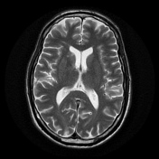
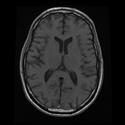
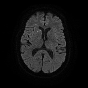
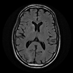
MRI 6 days post partially successful clot retrieval showing only small foci of right MCA territory infarction which is substantially smaller than the initial at risk brain seen on CT perfusion prior to clot removal. Unfortunately, the most prominent area of infarction is within the right pre-central gyrus at the hand bump and therefore likely to produce a neurological deficit.
Case Discussion
An example of a hyperacute MCA territory infarct with substantial ischemic penumbra determined on CT perfusion and good result post clot retrieval.
With thanks to Dr Anthony Kam and Dr Anoop Madan.


 Unable to process the form. Check for errors and try again.