
Presentation
Known knee osteoarthritis, now with severe pain and swelling in the left knee. No traumatic injury. Painful swelling and tenderness over the medial side of the knee and proximal tibia.
Patient Data
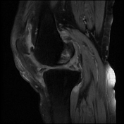

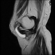
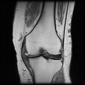
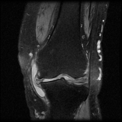
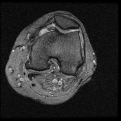
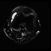

Elongated fluid collection interposed between the superficial tibial collateral ligament and the meniscofemoral and meniscotibial portions of the deep MCL. On axial images, the distended bursa has a lobulated appearance as it bulges slightly superficially around the anterior margin of the tibial collateral ligament.
Osteoarthritic changes in the knee with associated medial and lateral meniscal tears.
Impression
medial collateral bursitis
Case Discussion
The medial supporting structure of the knee can be separated into three layers. The first layer consists of the deep crural fascia. The superficial portion of the MCL is the main component of the second layer. The joint capsule and deep portion of the MCL which comprises the meniscotibial and meniscofemoral extensions make up the third layer.
The MCL bursa is located between the superficial and deep portions of the MCL and is not identified on MR imaging without a fluid collection.
MCL bursitis is infrequently seen but must be kept in mind for the differential diagnosis in patients with medial knee joint pain to avoid unnecessary arthroscopic surgery. Trauma, osteophytic spurs, rheumatic disorders, genu valgus, and flatfoot deformity have been suggested as causes of MCL bursitis.


 Unable to process the form. Check for errors and try again.