
Presentation
Knee pain.
Patient Data
Age: 45 years
Gender: Male
From the case:
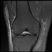
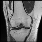
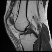
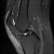
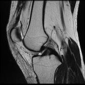
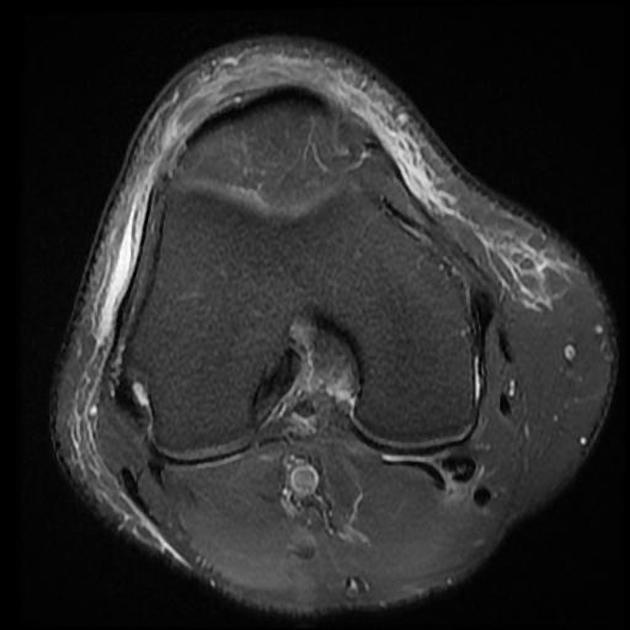
Medial discoid meniscus


Download
Info
Mild joint effusion.
Incomplete discoid medial meniscus with alteration of the signal due to intrameniscal mucoid degeneration.
The lateral meniscus seems discoid, with no abnormal signal identified to suggest the presence of a tear within.
Patella chondromalacia as cartilage thinning and fraying associated with subchondral cystic changes at the mid to medial facet joints of the patella.
Prepatellar bursitis and subcutaneous edematous changes along with mid to anterior aspect of the knee joint.
Case Discussion
Discoid menisci are those that have a body that is too wide. Usually, the lateral meniscus is affected, discoid medial menisci are rare.


 Unable to process the form. Check for errors and try again.