
Presentation
Tinnitus and vertigo
Patient Data
Age: 65 years
Gender: Male
From the case:
Ménière disease


Download
Info
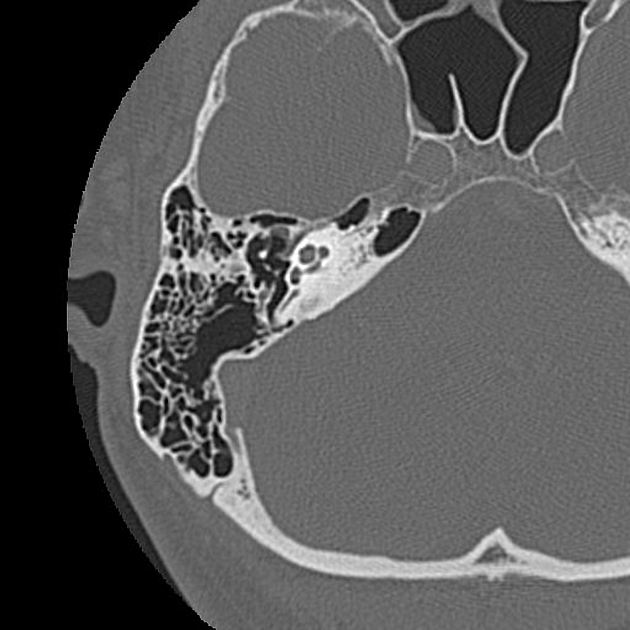
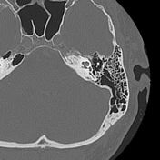
Endolymphatic ducts and sacs are faintly visualized bilaterally worse on the left. Vestibular aqueducts are also stenotic.
Case Discussion
Ménière disease is largely a clinical diagnosis but radiologic diagnosis can be achieved with high-resolution dedicated imaging of the petrous temporal bones. Both CT and MRI could be considered for any clinically suspected Ménière disease.


 Unable to process the form. Check for errors and try again.