
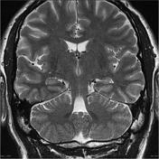

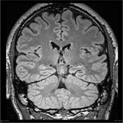

On T2, the left hippocampus (head and body) is smaller compared with right (see the apparent larger temporal horn). It has also lost the trilaminar appearance and undulating contour -- still preserved on the right. On FLAIR, there is subtle but definite increase in intensity within the left hippocampus.
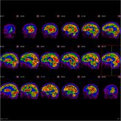
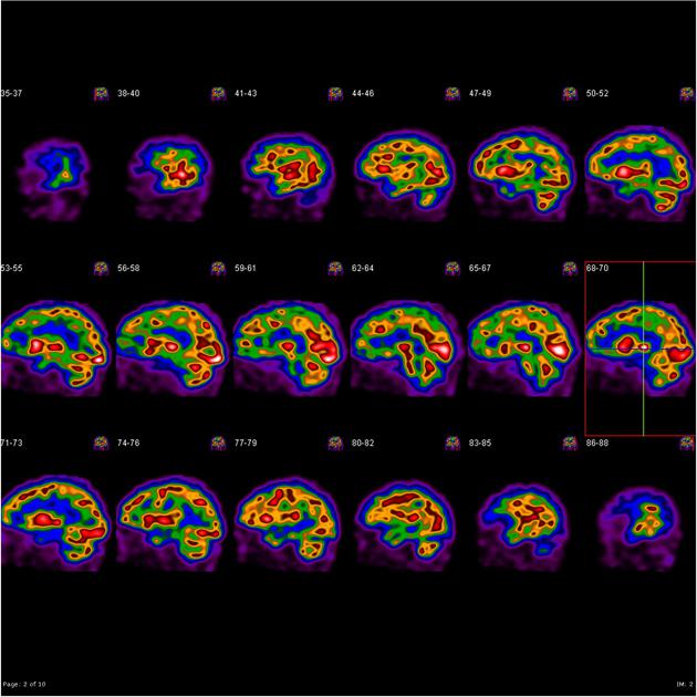
Ictal: No hyperperfusion seen. Hypoperfusion seen in left temporal and parietal lobes.
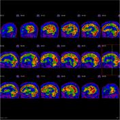
Interictal: Focal hypoperfusion of left temporal lobe.
Conclusion: Although there is no active focus of epilepsy, hypoperfusion of left temporal lobes in both studies is consistent with left MTS.
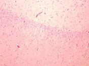

1. Temporal lobe lateral: Mild Chaslin's sclerosis only.
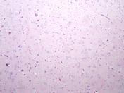
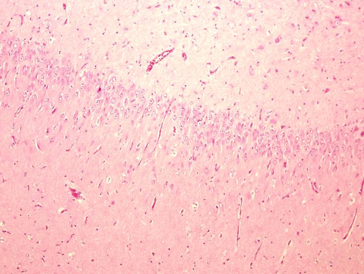
2. Hippocampus: Hippocampal sclerosis - Blumcke Type Ia.
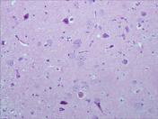
3. Maxilla, uncus; Focal cortical dysplasia - Taylor Type IIa.
Case Discussion
This case is in progress.


 Unable to process the form. Check for errors and try again.