Presentation
Complex partial seizures with déjà vu episodes, speech disturbance, and stereotyped motor episodes.
Patient Data
![](https://prod-images-static.radiopaedia.org/images/33366884/15850d3d806664739c99bd098a3b3c_thumb.jpeg)
![This study is a stack](/packs/stack-YQKLCKBI.gif)
![](https://prod-images-static.radiopaedia.org/images/33366863/422cdff4b09a1740c02f3767f0ffcd_thumb.jpeg)
![This study is a stack](/packs/stack-YQKLCKBI.gif)
![](https://prod-images-static.radiopaedia.org/images/33367071/1df9aa7b7b640dd03ad6486f585884_thumb.jpeg)
![This study is a stack](/packs/stack-YQKLCKBI.gif)
![](https://prod-images-static.radiopaedia.org/images/33366913/2301db65569b5a8f2a32ef05cd60d6_thumb.jpeg)
![This study is a stack](/packs/stack-YQKLCKBI.gif)
![](https://prod-images-static.radiopaedia.org/images/33368345/d13ef3a3f9c260b3070f27a9c8fb91_thumb.jpeg)
![This study is a stack](/packs/stack-YQKLCKBI.gif)
![](https://prod-images-static.radiopaedia.org/images/33366810/a90f57c5102827ee38c7a54138b94e_thumb.jpeg)
![This study is a stack](/packs/stack-YQKLCKBI.gif)
![](https://prod-images-static.radiopaedia.org/images/33366884/15850d3d806664739c99bd098a3b3c_big_gallery.jpeg)
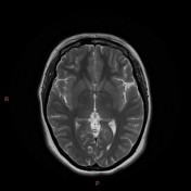
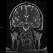
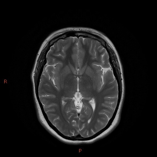
Small left hippocampal head and body with abnormal architecture, best demonstrated on the coronal T2W sequence. Subtle increased T2W/FLAIR signal.
No migrational abnormality or mass lesion.
![](https://prod-images-static.radiopaedia.org/images/33366850/177fc631414aa058cc905f38834893_thumb.jpeg)
![This study is a stack](/packs/stack-YQKLCKBI.gif)
![](https://prod-images-static.radiopaedia.org/images/33366954/28130f8e3a0d410a87b7e1eb61904a_thumb.jpeg)
![This study is a stack](/packs/stack-YQKLCKBI.gif)
![](https://prod-images-static.radiopaedia.org/images/33366850/177fc631414aa058cc905f38834893_big_gallery.jpeg)
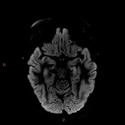
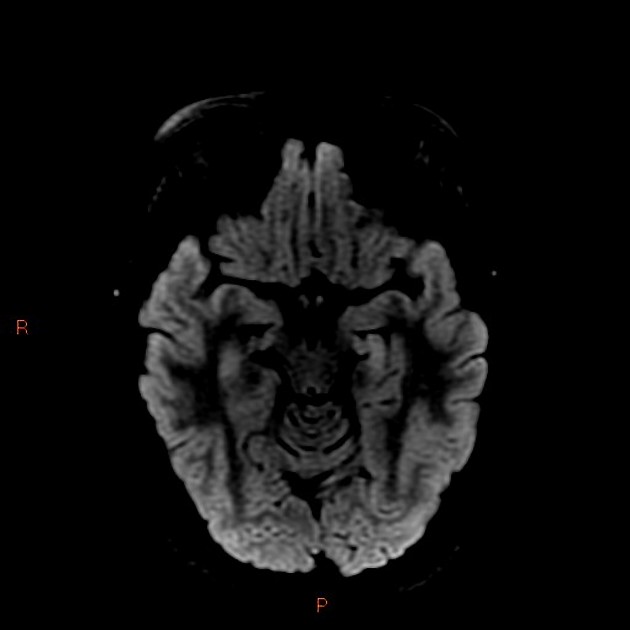
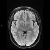
Focal increased FLAIR signal in the left hippocampus. On the coronal T2W sequence along the mesial temporal lobe the left collateral sulcus is too vertical in its alignment, consistent with incomplete inversion of the hippocampus.
![](https://prod-images-static.radiopaedia.org/images/33369432/e0bcc323671a9eb404157577c8695a_big_gallery.jpeg)
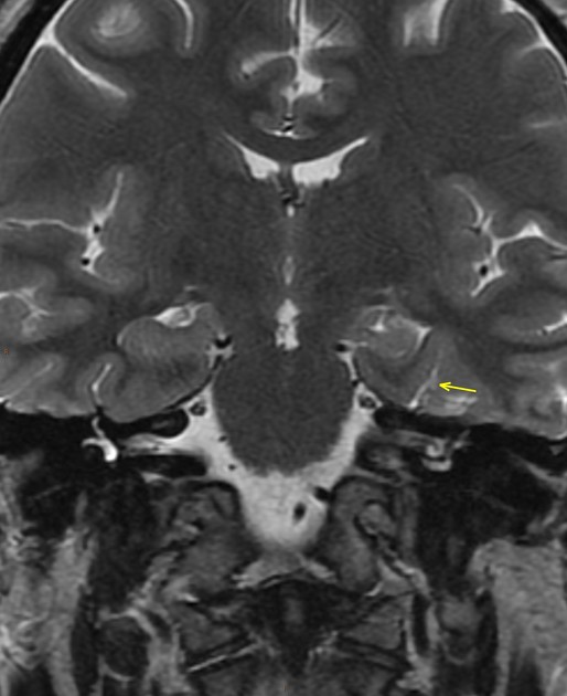
Zoomed and annotated coronal T2W sequence follows the left (yellow) and right (red) collateral sulci.
On the coronal T2W sequence along the mesial temporal lobe the left collateral sulcus is too vertical in its alignment, consistent with incomplete inversion of the hippocampus.
Case Discussion
Close inspection of the coronal T2W sequence shows not only atrophy and abnormal internal architecture of the left hippocampus, but also an abnormally vertical left collateral sulcus. This "incomplete inversion" of the hippocampus is in itself a potential epileptogenic focus.