
Presentation
Left submandibular swelling for last 5 months
Patient Data


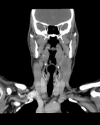
Septated conglomerate necrotic lymph nodes anterior to the left jugular vein, IIA level, measuring about 2.4 x 2.6 x 4 cm in AP, TS, and CC dimensions, compressing the jugular vein and pushing the left submandibular gland.
Other, multicompartmental enlarged cervical lymph nodes, the largest seen at the right mid jugular level, measuring about 1.4 cm in short axis.
Preservation of different neck space fat planes. Normal thyroid, parotid, and submandibular glands.
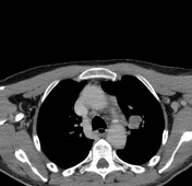
Multiple enlarged hypoattenuating left hilar lymph nodes are seen, the dominant node measures about 3.4 x 2.5 cm.
No evidence of lung masses, consolidation, or ILD.
No pericardial or pleural effusion. No pneumothorax.
No enlarged axillary lymph nodes.
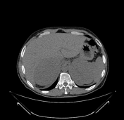
The visualized cuts of the abdomen show two large hypoattenuating lobulated masses at the site of the adrenal gland, displacing the abdominal organs and encasing vessels, measuring about 14 x 11 x 17.7 cm and 12 x 10 x 15 cm in AP, TS, and CC dimensions.
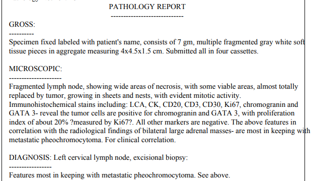
Case Discussion
The presence of local invasion or metastatic disease are the only ways to establish malignancy of pheochromocytoma.
Unfortunately the patient died before undergoing further imaging or investigations.


 Unable to process the form. Check for errors and try again.