
Presentation
Headache and unsteady gait for 5 days.
Patient Data
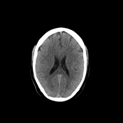

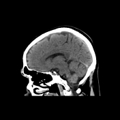
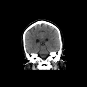

Non-contrast CT of the brain was unremarkable.
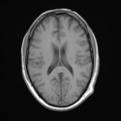
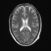


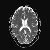
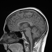
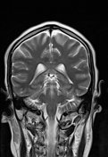

Bilateral symmetric T2 and FLAIR hyperintense lesions are seen in the dentate nuclei of the cerebellum, and the splenium of the corpus callosum. DWI shows mild hyperintensity of the splenium of the corpus callosum.
Case Discussion
The patient reports taking metronidazole for amoebiasis for the last 10 days.
Metronidazole-induced encephalopathy (MIE) always presents with bilateral and symmetrical hyperintense demyelinating lesions at the cerebellar dentate nuclei, dorsal medulla and pons, midbrain, and splenium of the corpus callosum. The lesions present with hyperintense T2 signal and best appreciated on FLAIR sequence. Interestingly, on DWI, most of the lesions in MIE do not show restricted diffusion as they would probably represent vasogenic edema, whereas only the corpus callosum lesions show mild restricted diffusion as they would correspond to cytotoxic edema.
Uncommonly, demyelinating lesions could be seen within the white matter of the cerebral hemispheres as well as the inferior olivary nucleus of the medulla.
The case is courtesy of Dr. Mohammed Ibrahim, MD.


 Unable to process the form. Check for errors and try again.