
Presentation
Hypertension.
Patient Data
Age: 20 years
Gender: Female
Note: This case has been tagged as "legacy" as it no longer meets image preparation and/or other case publication guidelines.
From the case:
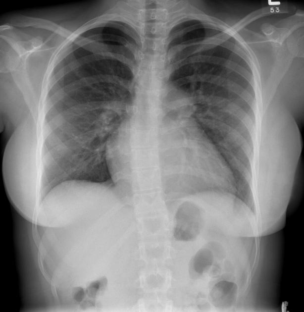
Midaortic syndrome

Download
Info

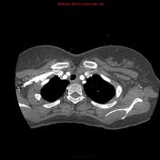
CT scan demonstrates narrowing of the descending aorta at the lower chest level, consistent with the diagnosis of midaortic syndrome.
A tight supravalvular pulmonary stenosis with post-stenotic dilatation is also seen.
From the case:
Midaortic syndrome
Download
Info
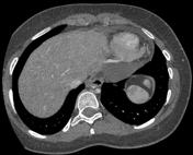
Severe left and moderate right ventricular hypertrophy are noted.
Case Discussion
Features of midaortic syndrome.


 Unable to process the form. Check for errors and try again.