
Presentation
A cardiomyopathy patient presents with a short history of right-sided paresis.
Patient Data
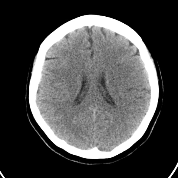
The M2-M3 segment of the left middle cerebral artery in the distal Sylvian fissure is hyperdense. It has a bright 'dot-like' like appearance on the axial images.
Little, if any, abnormality of distal brain parenchyma.
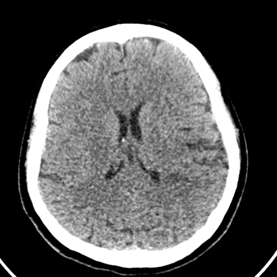
MRI abandoned due to lack of patient cooperation. 5 days later a rpt CTB was undertaken.
Relatively subtle low attenuation in the left temporoparietal region, in the distribution of the posterior branch of the MCA.
No hemorrhage.

Illustration of the anatomical arterial supply of the brain to illustrate the nature of the findings on the CT brain.
Thanks to Frank Gaillard for illustrations.
Case Discussion
A decent example of the MCA dot sign - the consequence of thrombus in the middle cerebral artery.
Subsequent end-organ damage in the form of a posterior branch MCA artery territory infarct is illustrated.


 Unable to process the form. Check for errors and try again.