
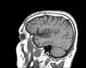
Presentation
Three days history of left hemiplegia.
Patient Data
Age: 50 years
Gender: Male
From the case:
Middle cerebral artery (MCA) infarct


Show annotations
Download
Info

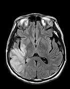
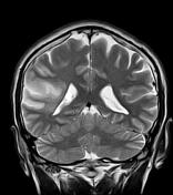
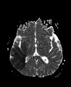
The MRI sequences demonstrate areas of low signal on T1, high signal on T2 and FLAIR with restricted diffusion in the distribution of the right middle cerebral artery (MCA) territory with no haemorrhagic transformation seen on the GE sequence.
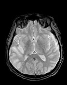
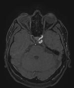
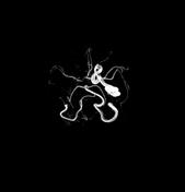
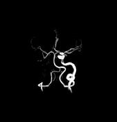
The MRA 3D-TOF shows reduced calibre of the right MCA and its branches with complete occlusion of the right internal carotid artery (ICA) well-demonstrated on MRA source images and 3D-TOF.
Case Discussion
MRI features of acute middle cerebral artery (MCA) territory infarct with complete occlusion of the internal carotid artery.


 Unable to process the form. Check for errors and try again.