
Presentation
Patient with autosomal dominant polycystic kidney disease presents with missing the intrauterine contraceptive device threads just after insertion two months ago.
Patient Data
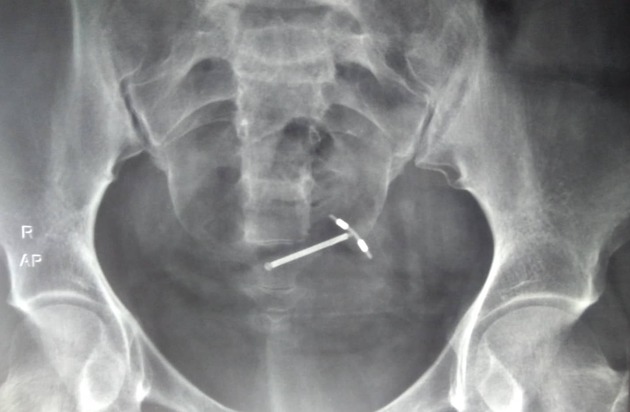
Pelvic radiograph showed the intrauterine contraceptive device lying obliquely within the true pelvis at the level of S4.
Patient was sent for transvaginal ultrasound for further verification.
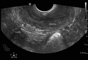
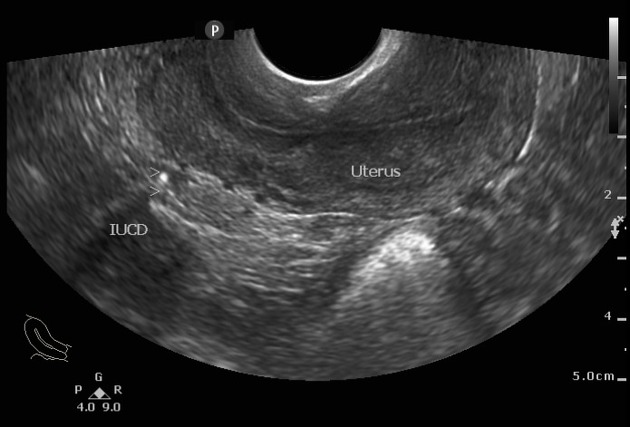
The uterus was shown empty along its cavity. The myometrium was intact with no gross abnormalities.
Longitudinally: an echoic pinpoint was noticed postero-superiorly a to the cervix (resembling the migrated contraceptive device).
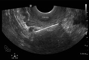
Transverse view showed contraceptive device within the Douglas pouch at the level of the cervix. Its full length was extruded out of the uterus.
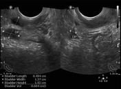
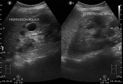
Three-dimensional measurement of the collection within the Douglas pouch showed insignificant amount of fluid (<7 mL). This tiny amount with the further abdominal scan of the Morrison pouch and the lienorenal space excluded the uncommon complication of significant hemoperitoneum.
The abdominal scan showed the previously reported ADPKD.
Case Discussion
Ultrasound is appropriate for initial evaluation and can provide answers to clinical questions related to the IUCD. The usual practice by the gynecologists to order abdomino-pelvic radiograph as an initial step should be reconsidered.
In this case, the radiograph alone could be misleading. The IUCD overlies the true pelvis with an acceptable orientation, yet the truth is that the IUCD lies outside the uterus. On the other hand, ultrasound saves unnecessary radiation exposure, accurately diagnoses the site and also detects complications.
However in cases with IUCD extruded to the abdominal cavity beyond the pelvis, radiography may have the upper hand.


 Unable to process the form. Check for errors and try again.